Visual Field Loss
WHAT IS FIELD LOSS?
Visual field loss is pretty much what it sounds like: the reduction in an area or extent of vision in one or both eyes. It encompasses a range of conditions, each with its own causes and treatments and each with its own types of vision loss. Understanding the type of vision field loss a patient is experiencing is crucial for figuring out the rest of the puzzle (the condition causing it and the appropriate treatment for it).
There are six main types of vision field loss:
- Central Vision Loss
- Peripheral Vision Loss
- Tunnel Vision
- Hemianopia
- Quadrantanopia
- Scotomas
What It Feels Like
Experiencing field loss can be unsettling and often frustrating. For many, it changes the way they interact with their surroundings and requires constant adjustments to navigate daily life. Depending on the type of field loss, individuals may experience:
Central Vision Loss
Central vision loss affects the central part of the visual field, which is the part necessary for activities like reading, driving, and recognizing faces. Patients with central vision loss often complain of a blurry or blank spot in their central vision.
Common causes include age-related macular degeneration (AMD), diabetic macular edema, and macular holes.
Treatments depend on the underlying cause and can range from injections and photodynamic therapy to overall lifestyle changes such as keeping blood sugar under control.
Peripheral Vision Loss
Peripheral vision loss affects the outer edges of the visual field but not the central vision. Because the vision loss isn’t directly in the middle of your view, patients suffering from peripheral vision loss are less likely to notice there’s an issue until it’s become fairly advanced. Unfortunately, early detection and treatment are crucial in treating this type of vision loss.
Causes include glaucoma, retinal detachment, and certain types of strokes.
Treatment for glaucoma includes medications to lower intraocular pressure, laser treatments, and surgery. For retinal detachment, surgical intervention is usually required.
Tunnel Vision
Tunnel vision is more than an accusation you fling at someone who can’t seem to see the forest from the trees. It’s a real thing. In fact, it’s a severe form of peripheral vision loss where the visual field is restricted to a small central area, making it feel like you’re looking through a tunnel.
It is commonly associated with advanced glaucoma, retinitis pigmentosa, and certain types of brain damage or stroke.
Management involves treating the underlying cause. For glaucoma, this includes medications and surgery. For retinitis pigmentosa, treatment is more supportive, focusing on maximizing remaining vision and using assistive technologies.
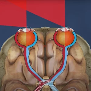
Hemianopia
Hemianopia is characterized by loss of vision in half of the visual field in one or both eyes. It can be homonymous (same half of the visual field in both eyes) or heteronymous (different halves in each eye).
This is commonly caused by brain injuries, strokes, or tumors affecting the visual pathways, and it often occurs when the occipital lobe or the optic tract are damaged.
While you can treat it with rehabilitation techniques such as visual field training, it’s crucial that you also treat the underlying cause (e.g., surgery or medication for tumors).
We have a whole section of our website dedicated to hemaniopia (AKA homonymous hemianopia) if you want to learn more about it and how we help.
Quadrantanopia
Quadrantanopia is the loss of vision in one-quarter of the visual field. This vision loss can occur in the upper or lower quadrants.
This type of vision loss is often the result of some type of brain damage (especially to the occipital lobe or optic radiation) from causes such as strokes or lesions in the brain.
The most useful way to treat this is via rehabilitation and compensatory techniques that help patients make use of their remaining vision and adapt to changes in their visual field.
Scotomas
Scotomas are specific areas of reduced or absent vision anywhere within the visual field. They can be any size, and they might present as blind spots or areas of blurred vision.
Causes include macular degeneration, diabetic retinopathy, and multiple sclerosis. Whether a scotoma is central or peripheral depends on the affected area of the retina or visual pathways.
Treatment depends on the cause. For conditions like macular degeneration, anti-VEGF injections or photodynamic therapy might be used. When it comes to preventing further scotomas from developing, managing conditions such as diabetes or multiple sclerosis is key.
Diagnostic Methods
Visual field loss is diagnosed via three different techniques:
- Visual Field Test
- Fundoscopy
- Imaging
A visual field test maps out the entire visual field and identifies any loss or abnormalities.
Fundoscopy is an examination that allows doctors to look at the retina and optic nerve for signs of disease.
Imaging includes techniques such as optical coherence tomography (OCT) or MRI, both of which can help visualize structural changes in the retina or brain.
General Treatments and Management
There are several ways to treat and/or manage visual field loss. The top 4 include:
Medical Treatment: Medications are often used to manage conditions like glaucoma and diabetic retinopathy. For inflammatory or infectious causes, specific medications or antibiotics might be prescribed.
Surgical Treatment: Surgery may be necessary for conditions like retinal detachment or advanced glaucoma. Laser therapy can also be used for certain retinal issues.
Rehabilitation: Vision therapy and rehabilitation can help patients adapt to vision field loss. Techniques might include training to improve the use of remaining vision, learning new visual skills, and using assistive devices.
Assistive Devices: Devices such as magnifiers, special glasses, and electronic visual aids can help individuals maximize their remaining vision and improve quality of life.
WHAT IS FIELD LOSS?
Visual field loss is pretty much what it sounds like: the reduction in an area or extent of vision in one or both eyes. It encompasses a range of conditions, each with its own causes and treatments and each with its own types of vision loss. Understanding the type of vision field loss a patient is experiencing is crucial for figuring out the rest of the puzzle (the condition causing it and the appropriate treatment for it).
There are six main types of vision field loss:
- Central Vision Loss
- Peripheral Vision Loss
- Tunnel Vision
- Hemianopia
- Quadrantanopia
- Scotomas
What It Feels Like
Experiencing field loss can be unsettling and often frustrating. For many, it changes the way they interact with their surroundings and requires constant adjustments to navigate daily life. Depending on the type of field loss, individuals may experience:
Central Vision Loss
Central vision loss affects the central part of the visual field, which is the part necessary for activities like reading, driving, and recognizing faces. Patients with central vision loss often complain of a blurry or blank spot in their central vision.
Common causes include age-related macular degeneration (AMD), diabetic macular edema, and macular holes.
Treatments depend on the underlying cause and can range from injections and photodynamic therapy to overall lifestyle changes such as keeping blood sugar under control.
Peripheral Vision Loss
Peripheral vision loss affects the outer edges of the visual field but not the central vision. Because the vision loss isn’t directly in the middle of your view, patients suffering from peripheral vision loss are less likely to notice there’s an issue until it’s become fairly advanced. Unfortunately, early detection and treatment are crucial in treating this type of vision loss.
Causes include glaucoma, retinal detachment, and certain types of strokes.
Treatment for glaucoma includes medications to lower intraocular pressure, laser treatments, and surgery. For retinal detachment, surgical intervention is usually required.
Tunnel Vision
Tunnel vision is more than an accusation you fling at someone who can’t seem to see the forest from the trees. It’s a real thing. In fact, it’s a severe form of peripheral vision loss where the visual field is restricted to a small central area, making it feel like you’re looking through a tunnel.
It is commonly associated with advanced glaucoma, retinitis pigmentosa, and certain types of brain damage or stroke.
Management involves treating the underlying cause. For glaucoma, this includes medications and surgery. For retinitis pigmentosa, treatment is more supportive, focusing on maximizing remaining vision and using assistive technologies.
Hemianopia
Hemianopia is characterized by loss of vision in half of the visual field in one or both eyes. It can be homonymous (same half of the visual field in both eyes) or heteronymous (different halves in each eye).
This is commonly caused by brain injuries, strokes, or tumors affecting the visual pathways, and it often occurs when the occipital lobe or the optic tract are damaged.
While you can treat it with rehabilitation techniques such as visual field training, it’s crucial that you also treat the underlying cause (e.g., surgery or medication for tumors).
We have a whole section of our website dedicated to hemaniopia (AKA homonymous hemianopia) if you want to learn more about it and how we help.
Quadrantanopia
Quadrantanopia is the loss of vision in one-quarter of the visual field. This vision loss can occur in the upper or lower quadrants.
This type of vision loss is often the result of some type of brain damage (especially to the occipital lobe or optic radiation) from causes such as strokes or lesions in the brain.
The most useful way to treat this is via rehabilitation and compensatory techniques that help patients make use of their remaining vision and adapt to changes in their visual field.
Scotomas
Scotomas are specific areas of reduced or absent vision anywhere within the visual field. They can be any size, and they might present as blind spots or areas of blurred vision.
Causes include macular degeneration, diabetic retinopathy, and multiple sclerosis. Whether a scotoma is central or peripheral depends on the affected area of the retina or visual pathways.
Treatment depends on the cause. For conditions like macular degeneration, anti-VEGF injections or photodynamic therapy might be used. When it comes to preventing further scotomas from developing, managing conditions such as diabetes or multiple sclerosis is key.
Diagnostic Methods
Visual field loss is diagnosed via three different techniques:
- Visual Field Test
- Fundoscopy
- Imaging
A visual field test maps out the entire visual field and identifies any loss or abnormalities.
Fundoscopy is an examination that allows doctors to look at the retina and optic nerve for signs of disease.
Imaging includes techniques such as optical coherence tomography (OCT) or MRI, both of which can help visualize structural changes in the retina or brain.
General Treatments and Management
There are several ways to treat and/or manage visual field loss. The top 4 include:
Medical Treatment: Medications are often used to manage conditions like glaucoma and diabetic retinopathy. For inflammatory or infectious causes, specific medications or antibiotics might be prescribed.
Surgical Treatment: Surgery may be necessary for conditions like retinal detachment or advanced glaucoma. Laser therapy can also be used for certain retinal issues.
Rehabilitation: Vision therapy and rehabilitation can help patients adapt to vision field loss. Techniques might include training to improve the use of remaining vision, learning new visual skills, and using assistive devices.
Assistive Devices: Devices such as magnifiers, special glasses, and electronic visual aids can help individuals maximize their remaining vision and improve quality of life.
How We Help
Central vision loss, peripheral vision loss, tunnel vision, hemianopia, quadrantanopia, and scotomas each present unique challenges. Diagnosing the specific type of visual field loss and understanding its cause are crucial for effective treatment and management. With advancements in medical and rehabilitative therapies, many individuals with vision field loss can improve their quality of life and maintain their independence
At Chadwick Optical, we have a variety of tools that can help individuals with field loss regain some functional vision and improve their ability to navigate their surroundings safely and confidently. Each person’s experience with field loss is unique, so our approach is grounded in offering a range of specialized devices designed to meet different types of field loss and to work alongside standard treatments and rehabilitation efforts.
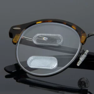
Peli Lens™
Prism lenses are an effective approach for managing certain types of field loss, particularly hemianopia (loss of half the visual field, often due to stroke). These lenses shift light from the area of field loss into the remaining functional visual field. The result? Users gain improved peripheral awareness, which can help with basic navigation and reducing the chances of bumping into obstacles.
High Powered Readers and Microscopic Spectacles
Our low vision aids are designed for individuals with significant field restrictions who require assistance with detailed tasks like reading, recognizing faces, or completing close-up work. This includes a range of high-magnification readers and microscopic spectacles. These aids can provide access to clearer, focused vision in a specific portion of the visual field, allowing patients to reclaim certain aspects of independence for daily tasks.
Microscopic spectacles provide extreme magnification for individuals with severely reduced vision, typically aiding in close-up tasks where precision is critical. Often used in cases where one eye has substantial impairment, these spectacles enhance vision in the other eye to compensate. These spectacles allow the user to engage in activities like reading fine print or inspecting small objects in detail.
Sunglasses and Tinted Lenses
For individuals with field loss due to light sensitivity or glare (often a secondary result of neurological conditions or eye diseases), we offer custom-tinted lenses and filters to manage discomfort and improve clarity. These specialized tints help patients to see contrasts and details more comfortably in bright environments, adding to the functionality of their remaining vision.